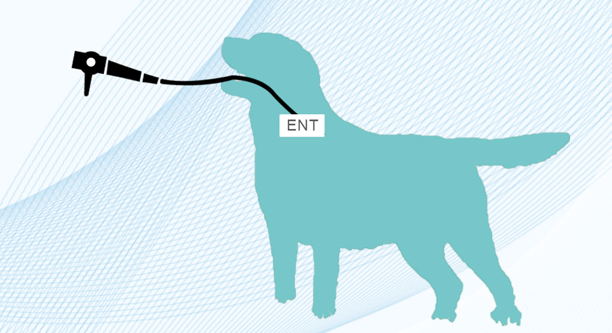
Endoscope technology has gradually become popular in pet hospitals. The use of endoscopes can provide a clearer mirror image of the pet's body, which can find problems and diseases, help the veterinarian to diagnose more accurately, and help the pet to recover in time. It is more common to take foreign objects. , The following endoscope manufacturers share the pet endoscope to take foreign objects, let's learn together.

The pet endoscope to take foreign bodies is that the pet endoscope can be used with professional tools to take out the foreign bodies swallowed by animals, which provides better skills for pet diagnosis and foreign body removal in the digestive tract.
Dogs are omnivores and often eat stones, plastic toys, food bags and other objects; cats usually eat linear substances, and linear foreign bodies are often entangled under the cat’s tongue or pylorus, causing intussusception. High-density substances (such as bones, fish hooks, etc.) can be diagnosed by X-rays, while some low-density substances (such as: rags, leather, etc.) are difficult to determine by X-rays, even if they are given barium preparations. It is difficult to determine the type, material, size, etc. of the foreign body, and the size and type of the foreign body in the digestive tract can be accurately diagnosed through the pet endoscope, and a more definite basis can be found for further treatment.
Operation process of pet endoscope:
The endoscope slowly passes through the larynx, advances slowly along the esophagus, and enters the stomach through the cardia. At the same time, there may be bubbles and food residues in the stomach. At this time, the electric suction device can be turned on for flushing and exhaust, and then the endoscope lens can be changed. The direction and the distance of advance and retreat, carefully observe and look for the suspicious foreign body in the stomach, until it is found, the foreign body can be grasped by the forceps with different functions, the size and nature of the foreign body can be confirmed, and some lesions can be collected for inspection, etc. Foreign body can try to use various functions of the clamp head to clamp the foreign body out of the body with the endoscope (such as: mouse tooth pliers, alligator pliers, three-jaw pliers, magnetic attraction rod, net blue, snare and outer tube, transparent Cap, etc.), to avoid the pain of animal surgery.
In some cases, surgery is required to retrieve foreign bodies:
Routinely open the abdominal cavity and find that the intestine is dilated due to gas. Find the abnormal intestine and find that the foreign body is located in the jejunum. Make an incision in the healthy intestinal tissue outside the distal end of the foreign body. Use a scalpel or surgery to cut the long axis of the intestine to open the incision to remove the foreign body. The intestine will not be torn. After removing the foreign body, trim the edge of the eversion mucosal layer to align with the serosal layer, butt the incision longitudinally or horizontally, and perform a simple intermittent suture across the entire thickness. The intestinal wall is fully sutured at a distance of 2mm from the edge. When the needle is fully penetrated, gently lift the needle tip and withdraw the needle until the mucosal layer slides out. Insert the needle from the contralateral mucosa-submucosa junction layer. Before inserting the needle, gently press the needle tip to turn the mucosal layer inward, and tighten carefully. Each suture, do not cut the intestinal wall layer, but gently but not crush the tissue.
Attention should be paid to foreign body removal during surgery:
1. Perform abdominal fluoroscopy or film before surgery to determine the location of the foreign body.
2. Insert a gastric tube before the operation to exhaust the contents of the stomach
3. The incision is determined according to the location of the foreign body. Whether it is a foreign body in the stomach or intestine, it is better to cut the gastrointestinal wall directly to remove the foreign body.
4. If the foreign body enters the duodenum and is accompanied by incarceration, it is best to push the foreign body into the stomach, and then cut a small opening in the stomach wall to take it out.
5. Take care to take all the foreign bodies out of the large number of foreign bodies. If possible, it is best to do an X-ray examination during the operation.
6. For those who have bleeding, perforation and peritonitis and other complications, in addition to taking foreign bodies, the complications should be treated accordingly.