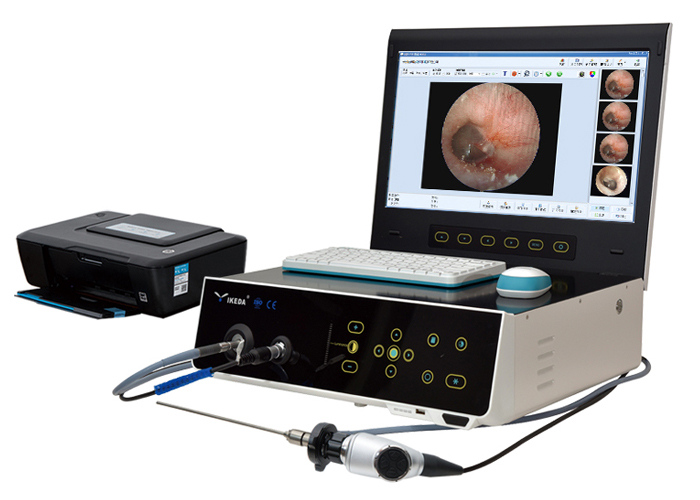
Endometrial cancer is a common malignant tumor of the female reproductive tract. Direct hysteroscopy and biopsy and pathological examination are the best methods for screening high-risk groups, early detection and accurate diagnosis of endometrial cancer and its precursors.
Indications for hysteroscopy:
1. Abnormal uterine bleeding;
2. Seen in abnormal sound image;
3. Infertility and family planning issues;
4. The physiological or special changes of the endometrium caused by hormone replacement or the application of tamoxifen.
Contraindications of hysteroscopy:
1. Pelvic infection;
2. Excessive uterine bleeding;
3. Those who want to continue pregnancy;
4. Recent uterine perforation;
5. The uterine cavity is too narrow or the cervix is too hard to dilate;
6. Those who suffer from serious medical illnesses who cannot tolerate uterine dilation operators;
7. Reproductive tract tuberculosis, without anti-tuberculosis treatment;
8. Those who have no follow-up treatment measures for blood diseases;
9. Invasive cervical cancer.
Hysteroscopy technology allows gynecologists to observe the entire uterine cavity at the most direct and closest distance without blind areas. Its small diameter and multifunctional design can perform endometrial positioning biopsy, especially the advent of fiber hysteroscopy, which can be applied to elderly women Diagnosis of intrauterine diseases. Hysteroscopy is simple in operation and accurate in diagnosis, and has become the "gold standard" for modern diagnosis of intrauterine lesions.
Main points of hysteroscopic diagnosis of endometrial cancer: Endometrial cancer may be seen when the following findings are present, and a biopsy must be sent for histopathological examination.
1. Translucent villous protuberances with mid-cardiovascular disease are likely to be well-differentiated endometrial adenocarcinoma.
2. There are abnormal blood vessels, especially dilated blood vessels with irregular shapes.
3. Nodular bulge or polyp bulge, fragile texture.
4. Necrotic tissues with white spots or spots.
The practicality of hysteroscopy:
1. Intimal tissues with varying degrees of toughness and abnormal blood vessels are highly suspected of new organisms.
2. From the special appearance of the bulge, it can sometimes be microscopically judged to be endometrial cancer.
3. For some lesions, the pathological tissue type or the degree of tissue differentiation can be inferred.
4. Determine the location of the lesion and make a direct biopsy, even if it is a small lesion, it can be correctly diagnosed, avoiding blind diagnosing and curettage.
5. Determine whether there is cancer infiltration in the cervical canal, and stage the endometrial cancer.
