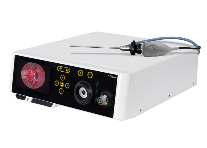
Through hysteroscopy, you can observe the cervical canal, the anterior and posterior walls of the uterus, the side walls, the bottom of the uterus, the left and right corners of the uterus and the opening of the fallopian tube to understand the shape of the uterine cavity and the endometrium. If necessary, perform corresponding diagnosing and curettage or Further other related surgical treatments were performed, and the intrauterine tissues were diagnosed and curettage sent for pathological examination.
Hysteroscopy procedure:
1. Dilate the cervix to the required size, place the hysteroscope into the cervix along the direction of the uterine cavity, and at the same time inject 5% glucose solution into the uterine cavity, rinse it out, and then inject glucose solution into the uterine cavity to dilate the uterus.
2. After the uterine cavity is fully expanded, you can use the hysteroscope to observe the morphology and endometrium of the uterine cavity, rotate the hysteroscope to sequentially check the various parts of the uterine cavity, and finally check the cervical canal, and then slowly withdraw from the cervix. During the inspection, the infusion of glucose solution must be maintained.
3. Through the operation pipeline of the official endoscopy, operations such as tissue biopsy and foreign body clamping can be performed.
What equipment does the hysteroscope include:
(1) Hysteroscope body: There are many types of hysteroscope, which can be basically divided into 4 types. ①Panoramic hysteroscope; ②Contact hysteroscope; ③Microscopic hysteroscope; ④Electric cutting hysteroscope.
(2) Hysteroscope tube sheath device.
(3) Hysteroscopic surgical instruments.
(4) Uterine dilatation device: 5% glucose solution is often used for dilatation liquid.
(5) Illumination system of the endoscope: including cooling light source and optical fiber.
(6) Endoscope camera system: optical converter, endoscope camera, high-definition monitor, workstation.
Hysteroscope is an advanced device for diagnosis and treatment of diseases in the uterine cavity. It can clearly observe various changes in the uterine cavity and make a clear diagnosis.
