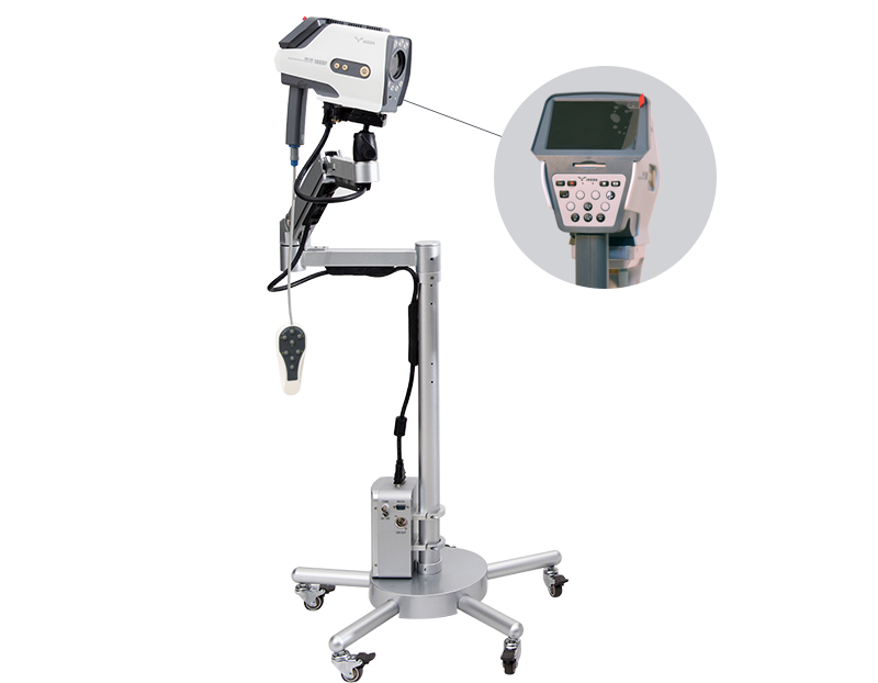
The digital video colposcopy system can magnify the uterine and vaginal mucosa, observe small changes in the cervical epidermis that are invisible to the naked eye, and discover abnormal epithelium and blood vessels related to cancer, so as to accurately select suspicious parts for biopsy.It is a powerful auxiliary method for diagnosing early cervical cancer.
How does the digital video colposcope system work?
When using the digital video colposcopy system, the doctor first enters the patient’s basic information, enters the personal information interface, and observes the image. The patient lies on the gynecological examination chair with the lens part 200~300 mm away from the patient. The LED cold light source will illuminate the patient’s vulva, In the vagina or cervix, the SONY color electronic imaging device COMS converts the collected optical signals into electrical signals, and converts the electrical signals into video signals through the control module, video capture card, and processing software, and displays them on the display. At the same time, they are adjusted and frozen by the control module. , Focus on the image signal, the footswitch collects video segments and pictures, the colposcopy software embeds the collected images into the software for display and storage, the physician combines the basic condition of the patient's examination and the examination results, analyzes and summarizes the diagnosis results in the form of graphic reports, and the printer prints color images The article reported that a biopsy of suspicious lesions was performed to further confirm the diagnosis.
The above briefly introduces How does the digital video colposcope system work. The digital video colposcope system is a gynecological examination and treatment system integrating photoelectric signals. It can diagnose and treat the abnormalities and diseased tissues of the vulva, vagina and cervix in time.
