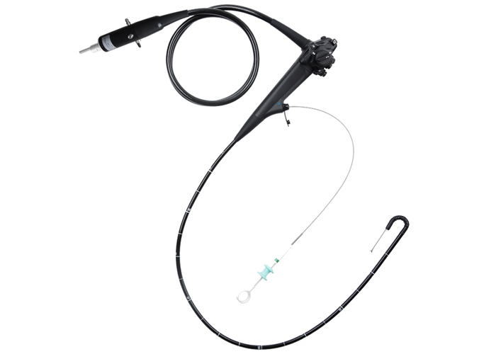
Common clinical applications of veterinary endoscopy include electronic gastroscope, veterinary laparoscope, arthroscope, otoscope, rhinoscope, cystoscope, etc. Electronic gastroscope is mainly used for visual inspection of esophagus, stomach and upper small intestine, etc. Get up and learn about veterinary electronic gastroscopy.

Electronic gastroscope indications:
Assess for dysphagia, gastroesophageal reflux, chronic vomiting, melena, chronic diarrhea, removal of esophageal foreign body, esophageal dilation, removal of part of gastric foreign body, and placement of gastric tube.
Contraindications: Contraindications to anesthesia, intestinal perforation, gastric accumulation, poor coagulation.
Common complications:
Digestive tract perforation, rupture of blood vessels, rupture of peripheral organs of gastrointestinal tract, acute bradycardia, insufficient venous return due to gastrointestinal gas, gastric torsion syndrome, mucosal hemorrhage, infection with Escherichia coli, bacteremia.
Types of electronic gastroscopes:
Rigid gastroscope: large in diameter, the advantage lies in grasping the foreign body in the esophagus, and sometimes the foreign body can be directly embedded in the lumen of the endoscope to prevent damage to the esophageal sphincter during the operation.
Flexible gastroscope: can be bent in four directions.
animal preparation
1. Fasting: at least 24h, do not feed dry food within 36h.
2. Anesthesia.
3. Baoding: lying on the left side of Baoding.
4. Install the mouthpiece, insert the endoscope, check while inserting, and control the air intake.
Examination of the esophagus:
The center of the esophagus remains in the center of the field of view, and when entering the esophagus through the annular pharyngeal sphincter, enough air is inhaled to dilate the esophagus.
normal:
The mucous membranes are pale and smooth.
Slightly rough behind the cricopharyngeal sphincter.
Submucosa vessels are clearly visible, especially near the sphincter at the lower end of the esophagus.
Stomach examination:
Once in the stomach, if there is no gas expansion (stomach contractions), a red smear is visible and the vision is unclear. At this time, the stomach should be inflated to expand the stomach to identify the gastric mucosa. The stomach is not full of gas, showing a collapsed state, prominent gastric folds, and gastric mucosa is rare; when the stomach is full of gas, most gastric folds disappear, and the mucosa can be increased.
Bend the mirror end as far back as possible to see the cardia. This field of view is best for gastric ulcers, and foreign bodies are sometimes found in this location. The end of the endoscope is pushed forward along the greater curvature of the stomach, and the end of the endoscope is slightly turned over.