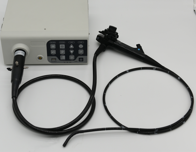
The electronic endoscope is mainly composed of three main parts: an endoscope, a TV information system and a TV monitor. It is also equipped with some auxiliary devices, such as a video recorder, a camera, a suction device, and a keyboard for inputting various information, as well as for diagnosis and treatment. Various disposal equipment, etc. Let us understand what is the imaging principle of electronic endoscopes? What is electronic endoscopy?
The imaging principle of electronic endoscope:
The optical fiber guides light into the cavity of the subject. The CCD image sensor receives the light reflected by the mucosal surface of the body cavity, converts this light into electrical signals, and transmits the signals to the TV information center through wires, and the TV information center passes these electrical signals through Storage and processing, and finally transmitted to the TV monitor to display the color image of the inspected organ on the screen.
What is the electronic endoscopy?
There are many types of electronic endoscopes on the market, and different electronic endoscopes have different functions. For example, gastroscopes can enter the stomach from the mouth and esophagus to check whether there are polyps, ulcers, and abnormal lesions in the stomach. Gastroscope can also be used for gastric mucosal polypectomy, needle biopsy of abnormal lesions, etc.
The electronic endoscope can detect the opening of the upper frontal sinus and the eustachian tube of the throat and whether there are any lesions in the lumen behind the ear. There is also a hysteroscope, which is used to check whether the uterus is deformed, whether the uterine wall is diseased, whether there is residual in the uterine cavity, etc., and can be operated in the uterine cavity.
Currently widely used electronic endoscopes, such as gastroscopy for gastric cancer; bronchoscopy for lung cancer and tracheal cancer; esophagoscopy for esophageal cancer; sigmoidoscopy for rectal cancer and sigmoid colon cancer; cystoscopy for bladder cancer; laryngoscopy for laryngeal cancer; Nasopharyngoscopy for nasopharyngeal cancer; hysteroscopy for cervical erosion, cervical cancer, and vaginal cancer.
Electronic gastrointestinal endoscopy operating procedures
1. Before endoscopy, make sure that the circuit, voltage and gastrointestinal endoscope performance are in good condition. Those undergoing gastroscopy should be prepared on an empty stomach, and those undergoing colonoscopy should be prepared to clean their intestines.
2. Introduce the necessity and safety of the examination to the patient before the examination, explain the essentials of cooperation and possible discomfort, so as to stabilize the patient's mood and obtain sufficient cooperation.
3. Fully understand the medical history, physical examination, barium meal, etc. before surgery to determine whether there are contraindications. Endoscopy should not be performed for patients with severe heart disease, hypertension, emphysema, or mental illness.
4. During the inspection, enter the mirror in strict accordance with the principle of "introduction through the cavity", observe while entering the mirror, and then carefully check when withdrawing the mirror. Observe each part of the stomach and each segment of the intestine in turn; find suspicious The lesion should be histological biopsy.
5. After the inspection of each case, exit the endoscope, thoroughly clean and disinfect according to the cleaning and disinfection procedures, in preparation for the next inspection.
6. After the inspection on the same day, thoroughly clean, disinfect, blow dry, and maintain the endoscope for later use.
